
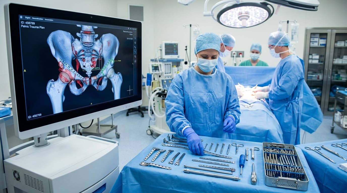

Understanding Pelvic & Acetabular Fracture Reconstruction
Pelvic ring injuries and acetabular (hip socket) fractures represent some of the most challenging conditions in orthopaedic trauma surgery. Typically caused by motor vehicle accidents, pedestrian collisions, or high falls, these injuries involve significant disruption of the body's central weight-bearing foundation and the delicate articular surface of the hip joint. Precise surgical reconstruction is essential to prevent permanent gait deformities, severe chronic pain, sciatic nerve compromise, and early-onset hip arthritis.
AIIMS Apex Trauma & AO Reconstruction Rigor
Complex articular fractures require anatomical joint surface restoration and stable biological fixation according to international AO principles to prevent post-traumatic arthritis and enable prompt joint mobilization.
When Should You Consider Consultation for Pelvic & Acetabular Surgery?
Specialist intervention is considered when joint pain or functional limitation significantly disrupts your daily life and conservative measures have been exhausted. Check if you experience any of the following:
High-energy motor vehicle accident, crush injury, or fall from height involving the pelvis or hip.
Severe, disabling pain in the groin, hip, or lower back with total inability to stand or bear weight.
Visible pelvic deformity, limb length discrepancy, or abnormal rotational position of the lower limb.
Secondary review of post-accident pelvic CT scans for complex acetabular column or wall fractures.
Second opinion on surgical indications for displaced acetabular fractures.
Management of persistent malunion or nonunion following prior conservative or emergency pelvic stabilization.

Dr. Vijay Kumar
Verified SpecialistSenior Consultant Orthopaedic, Robotic Joint Replacement & Complex Articular Trauma Surgeon
“My surgical philosophy centers on biological preservation, anatomical restoration, and rapid functional recovery. By tailoring the intervention to each patient's unique anatomy and lifestyle, we achieve lasting pain relief and active mobility.”
The 4-Stage Precision Clinical Workflow
3D CT Evaluation & Surgical Planning
High-resolution 3D CT volumetric reconstruction and Judet-Letournel classification mapping.
Direct Anatomical Articular Reduction
Selection of specialized surgical approaches (Kocher-Langenbeck, Ilioinguinal, Stoppa, or dual approaches) tailored to the fracture columns.
Rigid Internal Fixation (AO Protocol)
Direct anatomical reduction of the articular socket cartilage under direct optical visualization.
Supervised Early Mobilization & Healing
Rigid internal fixation using pre-contoured pelvic reconstruction plates and interfragmentary lag screws.
Stage 05: Reconstruction Stage
Intra-operative fluoroscopic verification of joint congruency and extra-articular hardware placement.
Treatment Modalities Offered
Dr. Vijay Kumar practices conservative joint-sparing protocols first, proceeding to advanced surgical intervention only when indicated:
Non-Surgical & Joint Sparing
- Non-operative treatment for strictly non-displaced pelvic fractures and stable pubic rami fractures
- Protected bed rest with sequential pneumatic compression to prevent deep vein thrombosis
- Strict non-weight-bearing mobilisation with walker under specialized physiotherapy supervision
- Serial radiographic monitoring to ensure fracture position remains stable
Specialised Surgical Procedures
- Open Reduction and Internal Fixation (ORIF) of Acetabular Fractures (Anterior & Posterior Columns)
- Anterior pelvic ring reconstruction using specialized anatomical curved pelvic plates
- Percutaneous sacroiliac (SI) screw fixation for posterior pelvic ring disruptions
- Combined Pelvic & Acetabular Reconstruction for complex multi-fragmentary fracture patterns
- Acute Total Hip Arthroplasty with acetabular reconstruction for elderly osteoporotic fracture-dislocations
Recovery Timeline & Mobility Milestones
Weeks 1–4: Gentle active-assisted hip range-of-motion exercises in bed; wheelchair or non-weight-bearing walker ambulation.
Weeks 6–8: Follow-up X-rays confirming early callus formation; initiation of partial weight bearing.
Weeks 10–14: Progressive full weight bearing, abductor muscle strengthening, weaning off crutches.
Months 4–6: Bone consolidation complete; resumption of active independent daily living and workplace duties.
International Fellowship & Surgical Suite Records
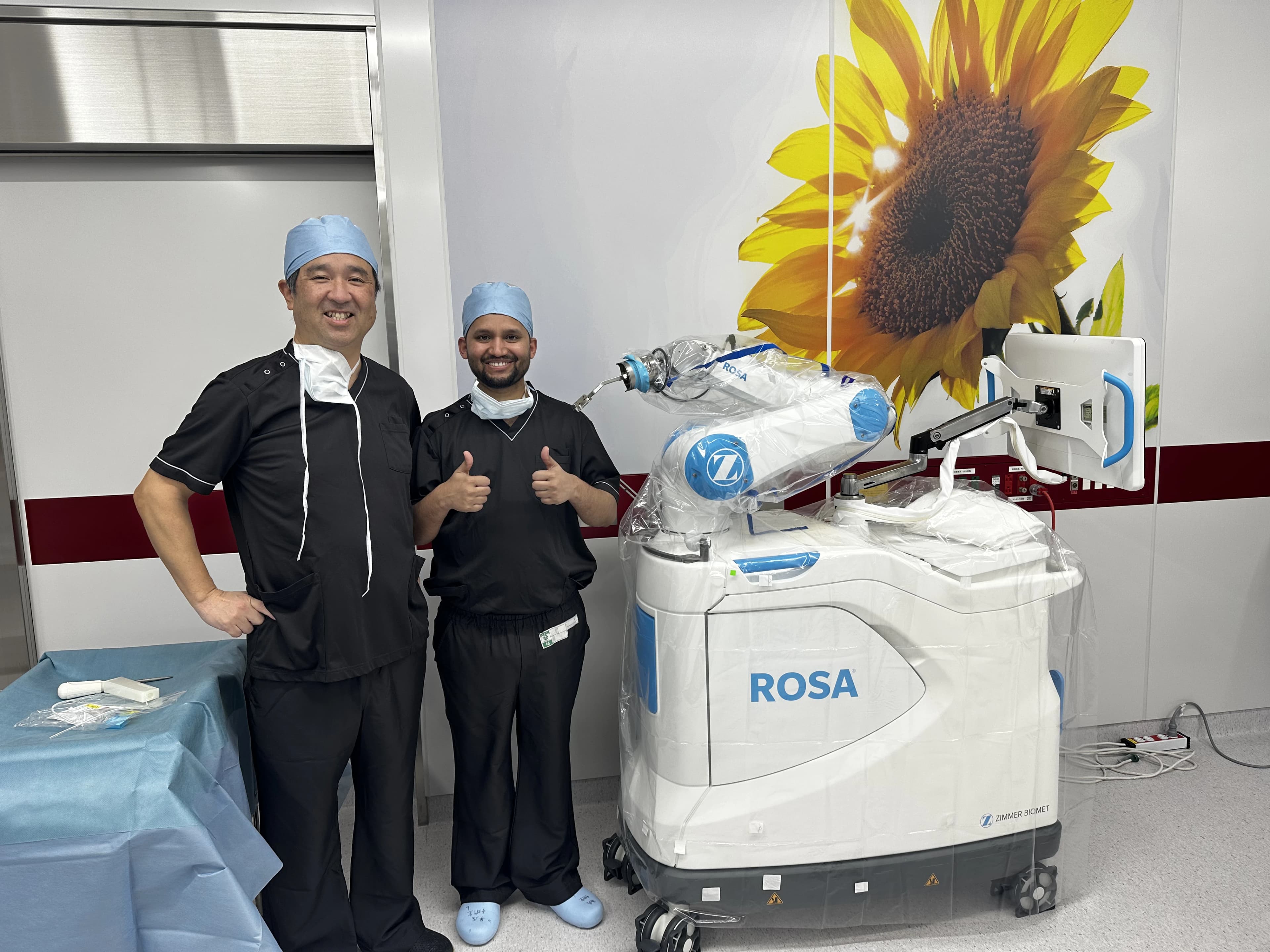
Intraoperative kinematics & 3D navigation alongside Japanese surgical faculty.
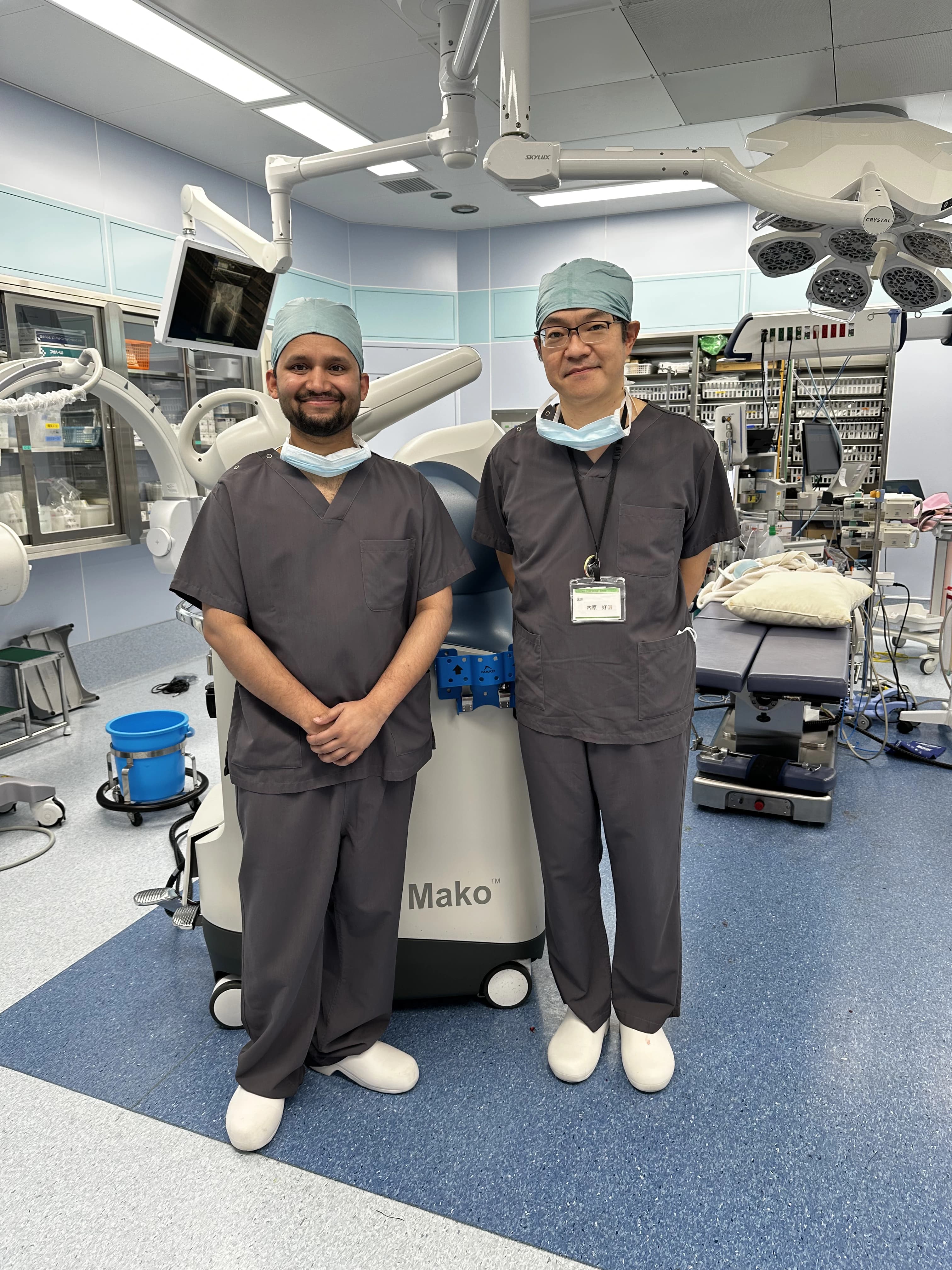
International fellowship-level precision applied to patient care in Patna.
Frequently Asked Questions on Pelvic & Acetabular Surgery
The acetabulum is the 'socket' of the ball-and-socket hip joint. When fractured with displacement, the joint surface becomes uneven. Walking on an uneven socket causes rapid cartilage wear and severe arthritis. Surgery restores a completely smooth socket surface to preserve your natural hip joint.