
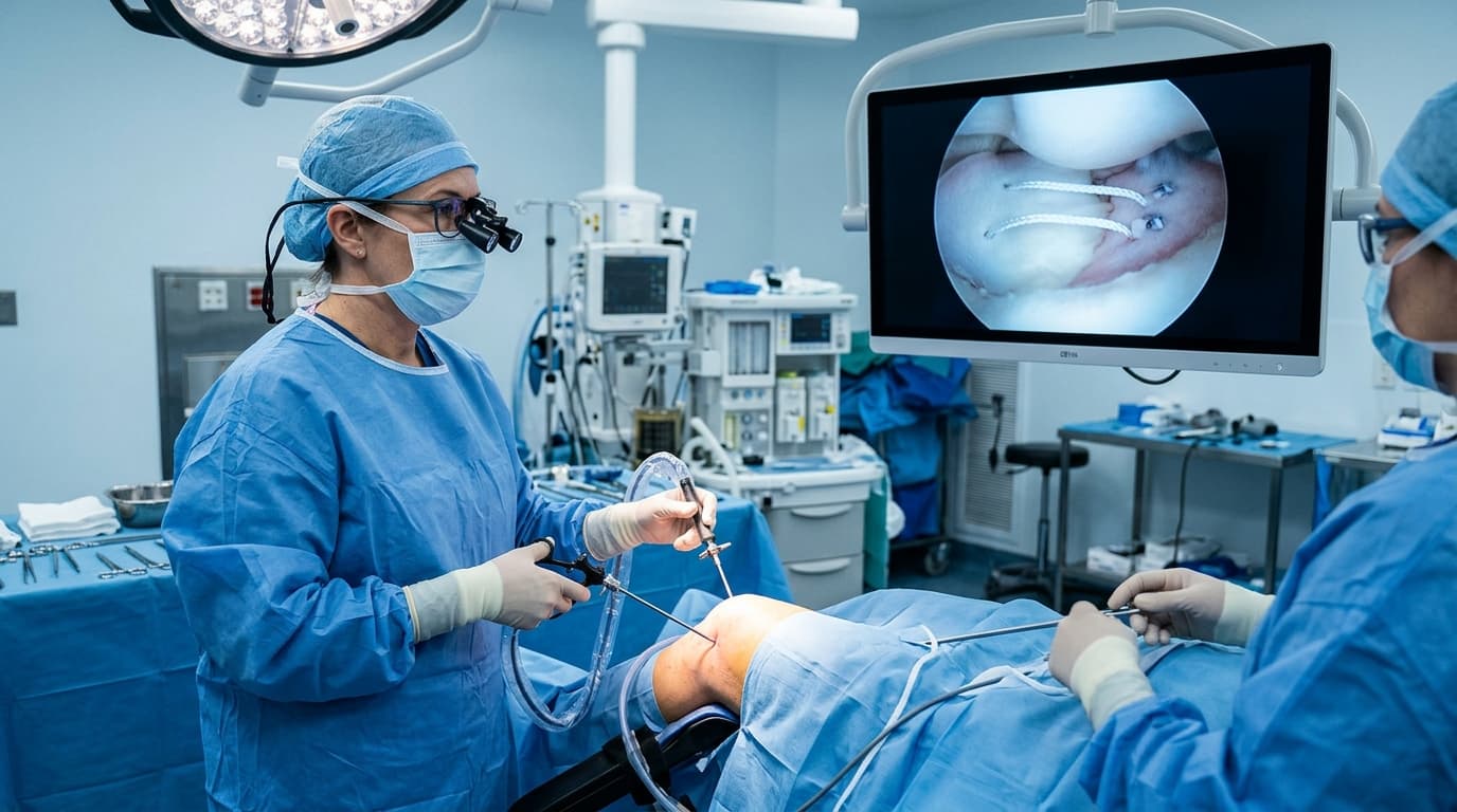

Understanding Meniscus Tear Repair & Advanced Meniscectomy
Meniscus tears occur either acutely in young athletes during sudden rotational twists or gradually in active adults due to age-related fibrocartilaginous wear. Symptoms include localized joint line pain, swelling, and mechanical catching or locking. Modern arthroscopic technique focuses on saving every millimeter of viable tissue through repair or performing selective partial contouring that removes only the torn, unstable flap while leaving the healthy peripheral shock-absorbing rim completely intact.
The Minimally Invasive Arthroscopy Advantage
Advanced 4K keyhole arthroscopy allows complete visualization and anatomical repair through pencil-thin incisions, eliminating extensive soft-tissue cutting, preserving natural joint biomechanics, and drastically accelerating recovery.
When Should You Consider Consultation for Meniscus Tear Care?
Specialist intervention is considered when joint pain or functional limitation significantly disrupts your daily life and conservative measures have been exhausted. Check if you experience any of the following:
Pain and localized tenderness on the inside or outside of the knee joint.
Knee catching, popping, or sudden inability to bend or straighten the leg (true locking).
Swelling that recurs after walking, running, or descending stairs.
Failure of rest, physiotherapy, or anti-inflammatory medications to resolve mechanical symptoms.
MRI diagnosis of a complex, horizontal, radial, or degenerative flap tear.

Dr. Vijay Kumar
Verified SpecialistSenior Consultant Orthopaedic, Robotic Joint Replacement & Complex Articular Trauma Surgeon
“My surgical philosophy centers on biological preservation, anatomical restoration, and rapid functional recovery. By tailoring the intervention to each patient's unique anatomy and lifestyle, we achieve lasting pain relief and active mobility.”
The 4-Stage Precision Clinical Workflow
Pre-Operative MRI & Diagnostic Mapping
4K optical insertion through two tiny 4-mm puncture portals.
High-Definition 4K Arthroscopic Access
Systematic probing of the meniscus upper and lower surfaces determining tissue stability.
Precision Anatomical Repair / Reconstruction
Precision resection of only unstable torn fragments using miniature motorized instruments.
Bio-Fixation & Sports Rehab Roadmap
Smoothing of transition zones to prevent secondary propagation of tears.
Stage 05: Arthroscopic Execution
Immediate joint irrigation and closure with minimal adhesive strips.
Treatment Modalities Offered
Dr. Vijay Kumar practices conservative joint-sparing protocols first, proceeding to advanced surgical intervention only when indicated:
Non-Surgical & Joint Sparing
- Structured quadriceps and hamstring strengthening therapy
- Activity modification avoiding deep squatting and pivoting
- Temporary non-steroidal anti-inflammatory medications
- Platelet-Rich Plasma (PRP) injection for minor intrameniscal signal changes
Specialised Surgical Procedures
- All-Inside and Inside-Out High-Tensile Meniscus Suture Repair
- Selective Arthroscopic Partial Meniscectomy (trimming unstable flap only)
- Meniscal Cyst Decompression and Excision
- Fibrin Clot Application to stimulate biological healing
Recovery Timeline & Mobility Milestones
Days 1–3: Immediate full weight bearing with or without single crutch, swelling control.
Weeks 1–2: Normal walking, full active range of motion, return to work.
Weeks 3–4: Stationary cycling, swimming, resistance band strengthening.
Weeks 4–6: Unrestricted sports, running, and athletic play.
International Fellowship & Surgical Suite Records
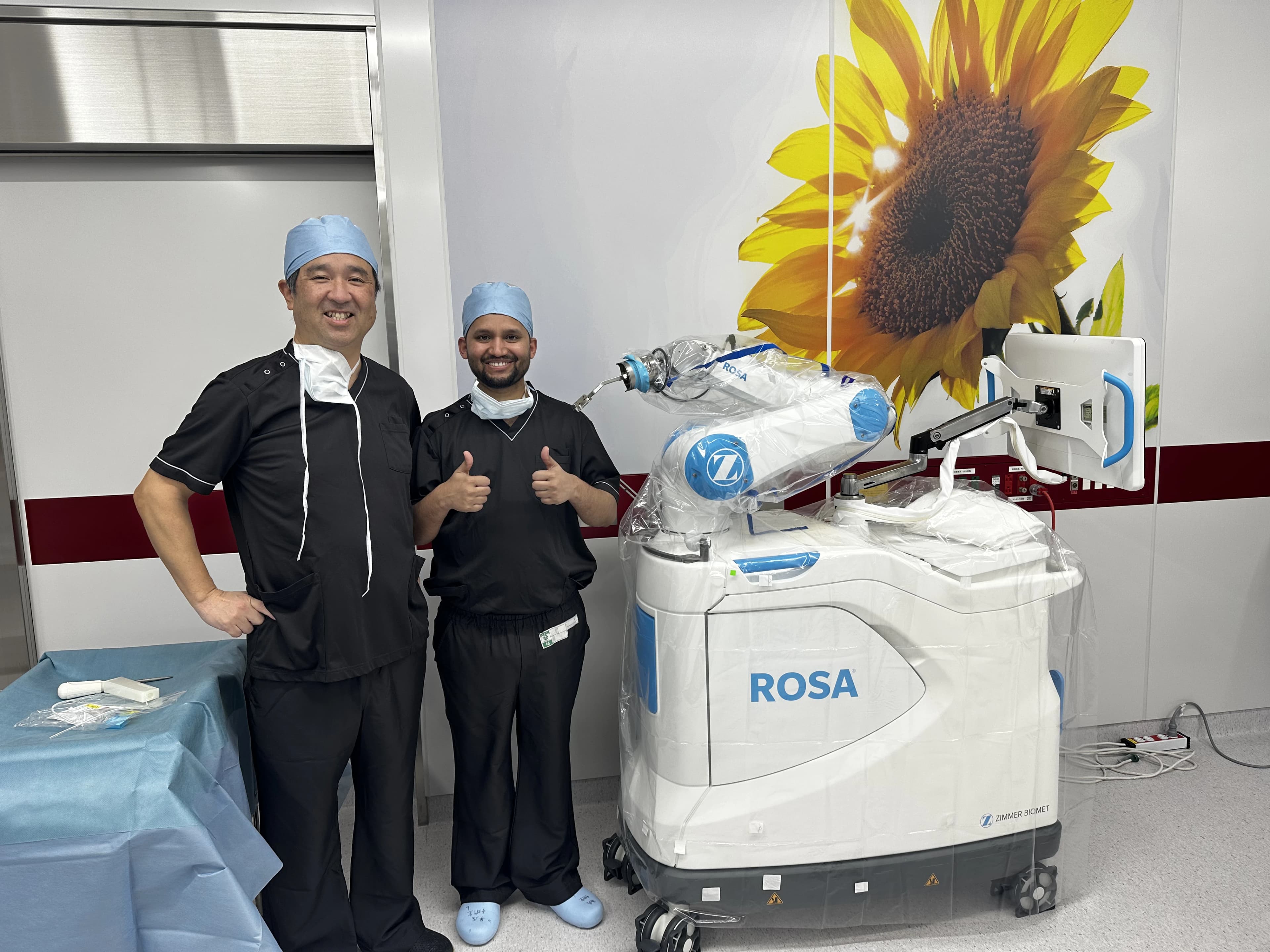
Intraoperative kinematics & 3D navigation alongside Japanese surgical faculty.
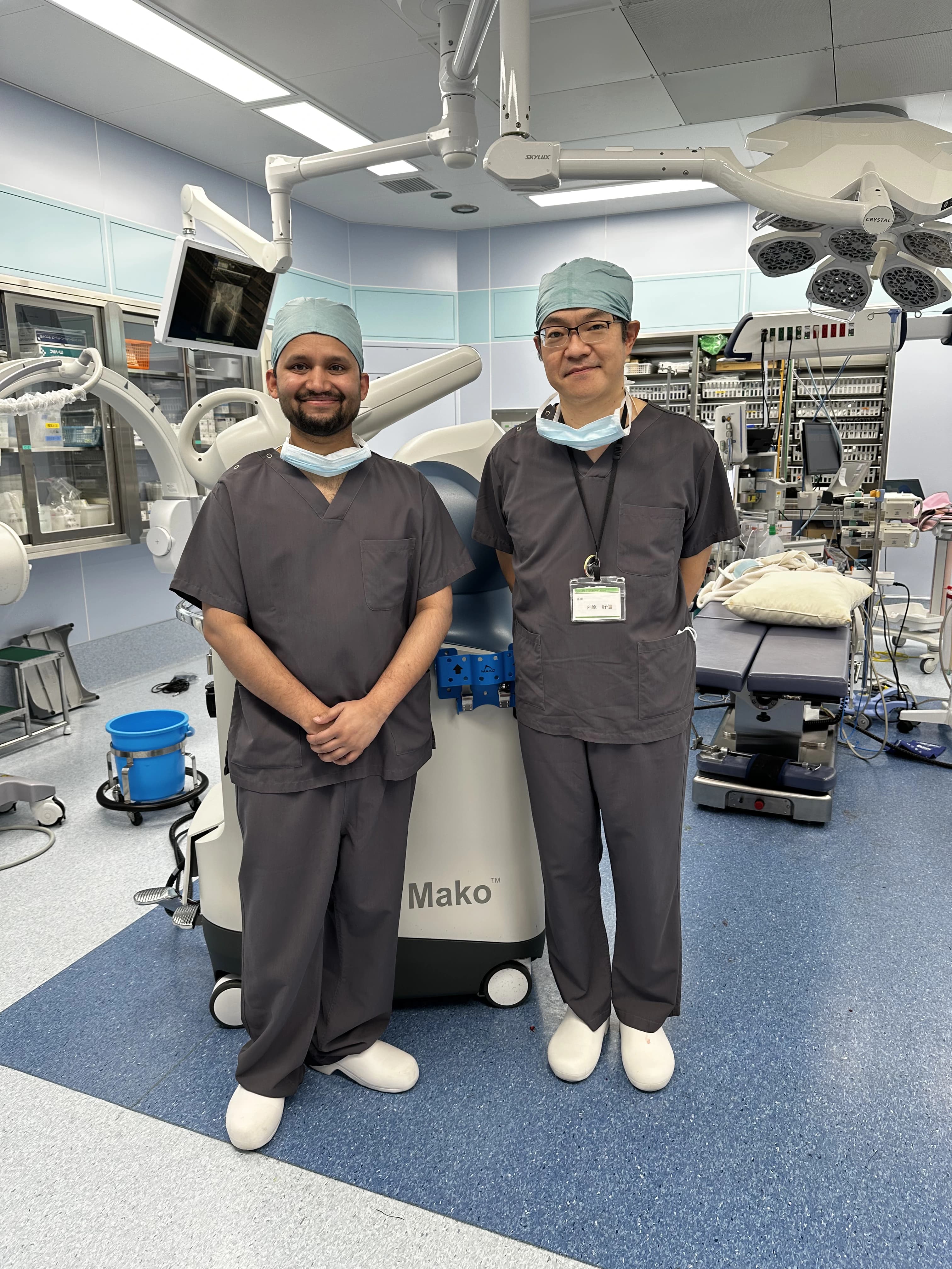
International fellowship-level precision applied to patient care in Patna.
Frequently Asked Questions on Meniscus Tear Care
Following a partial meniscectomy, patients walk with full weight bearing on the same day and typically return to light daily activities within 3 to 5 days.