

Understanding Hip Arthroscopy (FAI, Labral Tears & Synovitis)
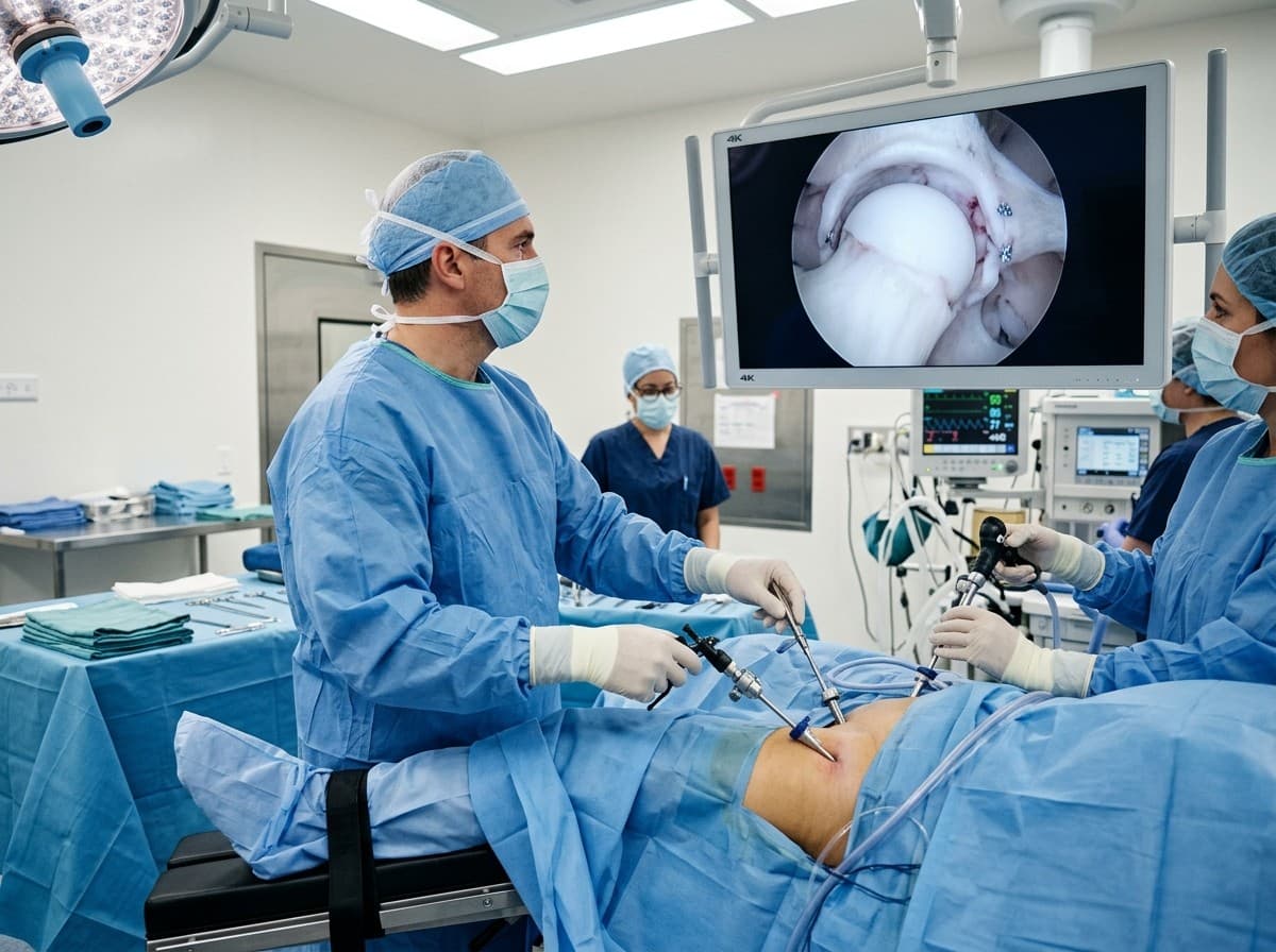
Femoroacetabular Impingement (FAI) is a condition where abnormal bone contours on the femoral head (Cam lesion) or acetabular socket (Pincer lesion) cause repetitive friction during hip flexion and rotation. This friction pinches and tears the acetabular labrum—the fibrocartilaginous seal of the hip—and damages surrounding articular cartilage, causing sharp groin pain during sitting, driving, or sports. 4K hip arthroscopy re-attaches the torn labrum with bio-anchors and reshapes the bone contours to restore smooth, friction-free movement.
The Minimally Invasive Arthroscopy Advantage
Advanced 4K keyhole arthroscopy allows complete visualization and anatomical repair through pencil-thin incisions, eliminating extensive soft-tissue cutting, preserving natural joint biomechanics, and drastically accelerating recovery.
When Should You Consider Consultation for Hip Arthroscopy (FAI)?
Specialist intervention is considered when joint pain or functional limitation significantly disrupts your daily life and conservative measures have been exhausted. Check if you experience any of the following:
Deep, aching groin or outer hip pain that worsens with prolonged sitting, driving, or hip flexion.
Sharp 'C-sign' pain in the hip during athletic pivoting, squatting, or martial arts.
Clicking, snapping, or catching sensations deep within the hip joint.
Young athlete or active individual diagnosed with FAI or labral tear on high-resolution MRI / 3T arthrogram.
Lack of lasting relief from physiotherapy, activity modification, or ultrasound-guided hip injections.

Dr. Vijay Kumar
Verified SpecialistSenior Consultant Orthopaedic, Robotic Joint Replacement & Complex Articular Trauma Surgeon
“My surgical philosophy centers on biological preservation, anatomical restoration, and rapid functional recovery. By tailoring the intervention to each patient's unique anatomy and lifestyle, we achieve lasting pain relief and active mobility.”
The 4-Stage Precision Clinical Workflow
Pre-Operative MRI & Diagnostic Mapping
Specialized traction table setup allowing safe access into the central hip joint under fluoroscopy.
High-Definition 4K Arthroscopic Access
4K optical visualization of the acetabular labrum, cartilage, and femoral neck.
Precision Anatomical Repair / Reconstruction
Placement of miniature bio-composite suture anchors securing the torn labral seal back to the socket.
Bio-Fixation & Sports Rehab Roadmap
Motorized burr recontouring of the Cam/Pincer bone spurs creating smooth clearance.
Stage 05: Arthroscopic Execution
Watertight capsular closure to maintain normal joint stability.
Treatment Modalities Offered
Dr. Vijay Kumar practices conservative joint-sparing protocols first, proceeding to advanced surgical intervention only when indicated:
Non-Surgical & Joint Sparing
- Specialized hip core and gluteal stabilizing physiotherapy
- Postural modification avoiding deep hip flexion past 90°
- Non-steroidal anti-inflammatory therapy for acute synovitis
- Ultrasound-guided diagnostic and therapeutic intra-articular hip injections
Specialised Surgical Procedures
- All-Arthroscopic Acetabular Labral Repair with knotless suture anchors
- Arthroscopic Cam Reshaping (Femoroplasty) and Pincer Trimming (Acetabuloplasty)
- Capsular Plication to restore joint stability in hypermobile patients
- Subchondral Microfracture / AMIC for focal chondral defects
Recovery Timeline & Mobility Milestones
Weeks 1–3: Crutch-assisted walking, protected hip flexion (<90°), circumduction exercises.
Weeks 4–6: Weaning off crutches, full weight bearing, progressive core and abductor conditioning.
Months 2–3: Elliptical, swimming, resistance band hip strengthening.
Months 4–6: Sport-specific rotational drills, sprinting, and clearance for contact athletics.
International Fellowship & Surgical Suite Records
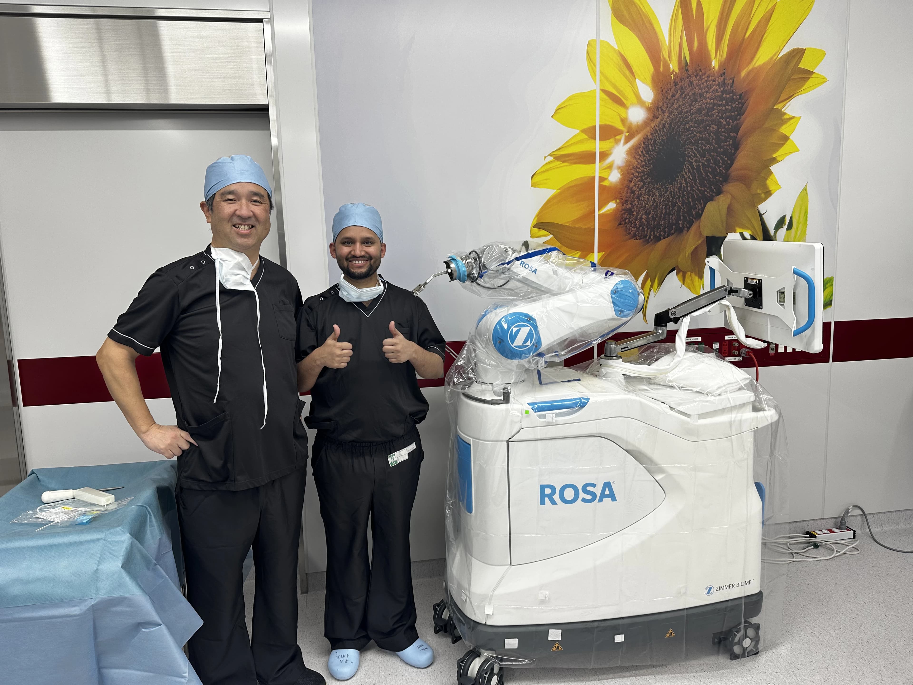
Intraoperative kinematics & 3D navigation alongside Japanese surgical faculty.
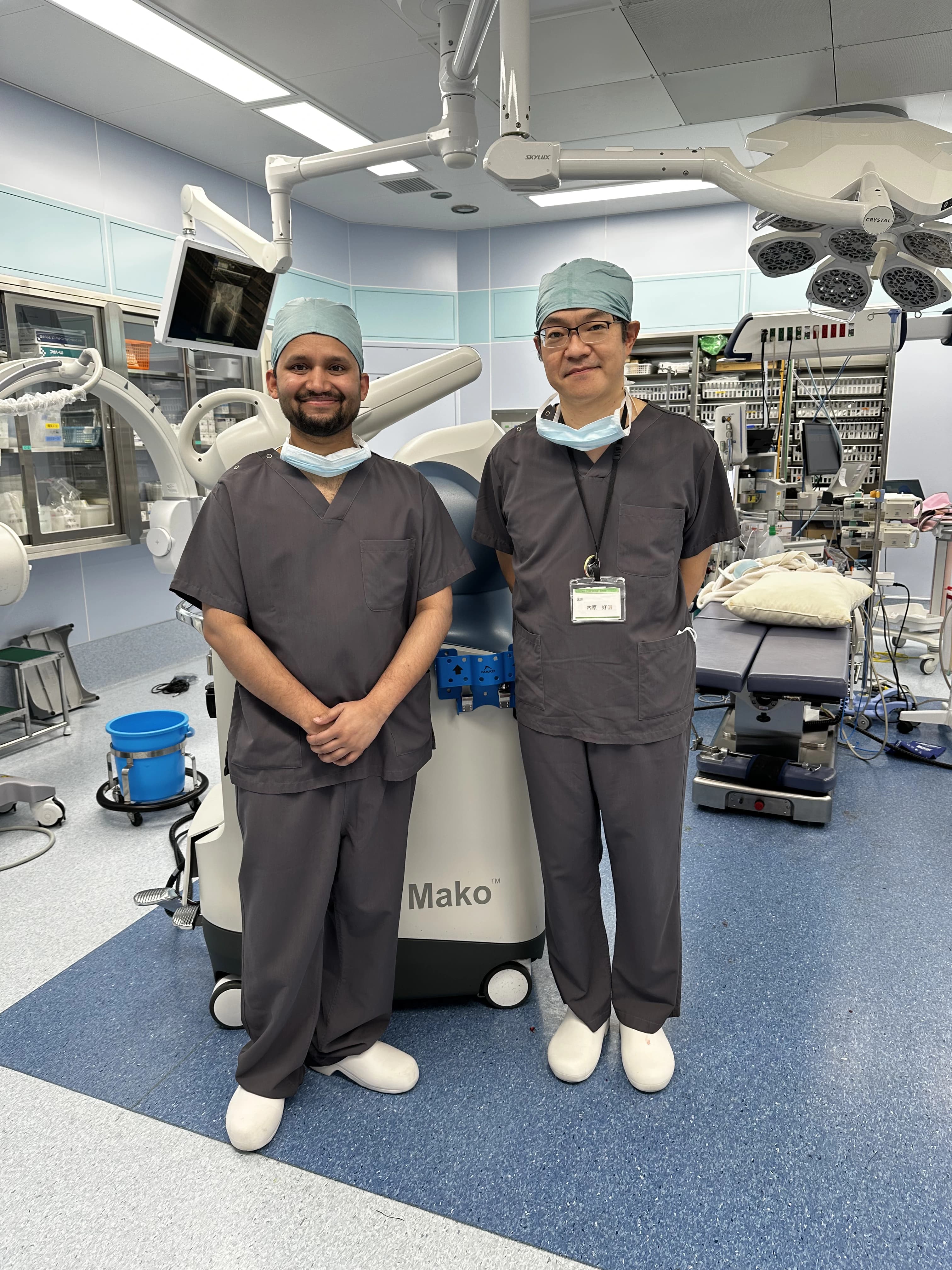
International fellowship-level precision applied to patient care in Patna.
Frequently Asked Questions on Hip Arthroscopy (FAI)
Yes. Untreated bone impingement continually shears the labrum and cartilage, leading directly to premature hip osteoarthritis in young and middle-aged adults. Arthroscopic correction eliminates impingement and protects the joint.