
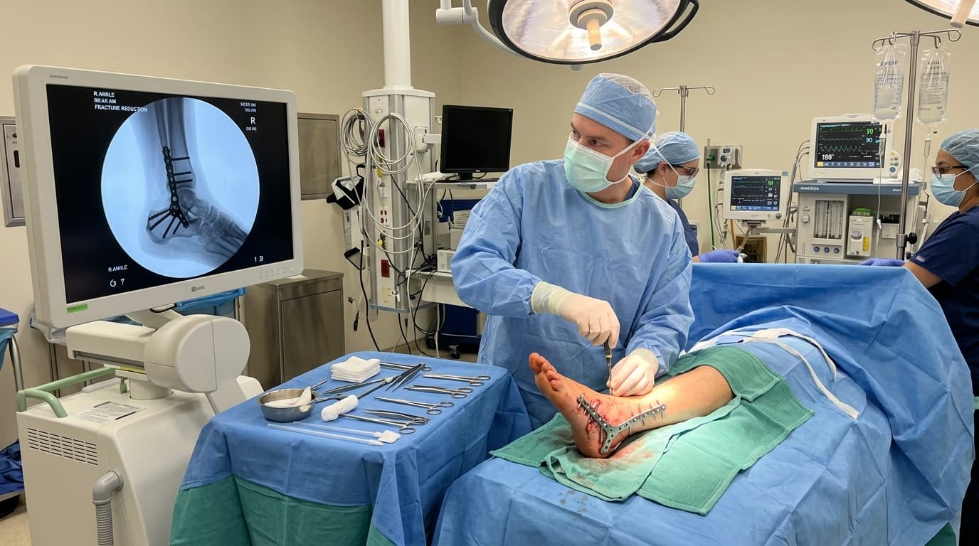

Understanding Foot & Ankle Fractures & Complex Calcaneal Trauma
The foot and ankle complex bears the highest load per square centimeter of any joint in the human body. High-energy twisting, motor vehicle crashes, or falls from ladders can cause complex fractures—including bimalleolar/trimalleolar ankle fractures, Pilon fractures, intra-articular calcaneal (heel) crushes, and Lisfranc midfoot ligament disruptions. Because even a 1-millimeter displacement of the talus inside the ankle socket reduces joint contact area by over 40%, anatomical surgical reduction and rigid low-profile plating are essential to prevent debilitating post-traumatic arthritis.
AIIMS Apex Trauma & AO Reconstruction Rigor
Complex articular fractures require anatomical joint surface restoration and stable biological fixation according to international AO principles to prevent post-traumatic arthritis and enable prompt joint mobilization.
When Should You Consider Consultation for Foot & Ankle Fractures?
Specialist intervention is considered when joint pain or functional limitation significantly disrupts your daily life and conservative measures have been exhausted. Check if you experience any of the following:
Severe ankle or foot pain, swelling, bruising, and inability to bear weight after a twisting fall or accident.
Visible deformity, skin tenting, or abnormal alignment of the ankle or foot.
Heel pain and flattening of the foot arch following a jump or fall from height (Calcaneus fracture).
Pain across the midfoot arch with plantar bruising after a direct crush or twisting stumble (Lisfranc injury).
Secondary review of complex post-accident foot and ankle CT scans.

Dr. Vijay Kumar
Verified SpecialistSenior Consultant Orthopaedic, Robotic Joint Replacement & Complex Articular Trauma Surgeon
“My surgical philosophy centers on biological preservation, anatomical restoration, and rapid functional recovery. By tailoring the intervention to each patient's unique anatomy and lifestyle, we achieve lasting pain relief and active mobility.”
The 4-Stage Precision Clinical Workflow
3D CT Evaluation & Surgical Planning
High-resolution 3D CT reconstruction analyzing articular joint depression and comminution.
Direct Anatomical Articular Reduction
Cosmetic, soft-tissue-preserving surgical approaches minimizing wound tension.
Rigid Internal Fixation (AO Protocol)
Direct anatomical reduction of the articular surface under direct optical view and fluoroscopy.
Supervised Early Mobilization & Healing
Rigid fixation using pre-contoured low-profile titanium locking plates and lag screws.
Stage 05: Reconstruction Stage
Application of a lightweight splint with early elevation to rapidly reduce swelling.
Treatment Modalities Offered
Dr. Vijay Kumar practices conservative joint-sparing protocols first, proceeding to advanced surgical intervention only when indicated:
Non-Surgical & Joint Sparing
- Non-weight-bearing cast immobilization for non-displaced, stable ankle fractures
- Removable pneumatic walking boot following initial bone union
- Serial radiographic monitoring to confirm stable alignment
Specialised Surgical Procedures
- Open Reduction and Internal Fixation (ORIF) for bimalleolar and trimalleolar ankle fractures
- Minimally Invasive Sinus Tarsi Approach for Calcaneus Fractures
- Lisfranc Joint Anatomical Screw / Bridge Plate Fixation
- Syndesmosis Suture-Button Dynamic Stabilization (TightRope)
- Anatomical Talar Neck and Body Reconstruction
Recovery Timeline & Mobility Milestones
Weeks 1–2: Strict foot elevation, swelling control, gentle toe movement.
Weeks 3–6: Non-weight-bearing walker ambulation, active ankle range of motion exercises.
Weeks 6–10: Follow-up X-rays confirming solid bone union; progressive full weight-bearing in supportive boot.
Months 3–4: Transition to standard athletic footwear, normal walking, return to light jogging.
International Fellowship & Surgical Suite Records
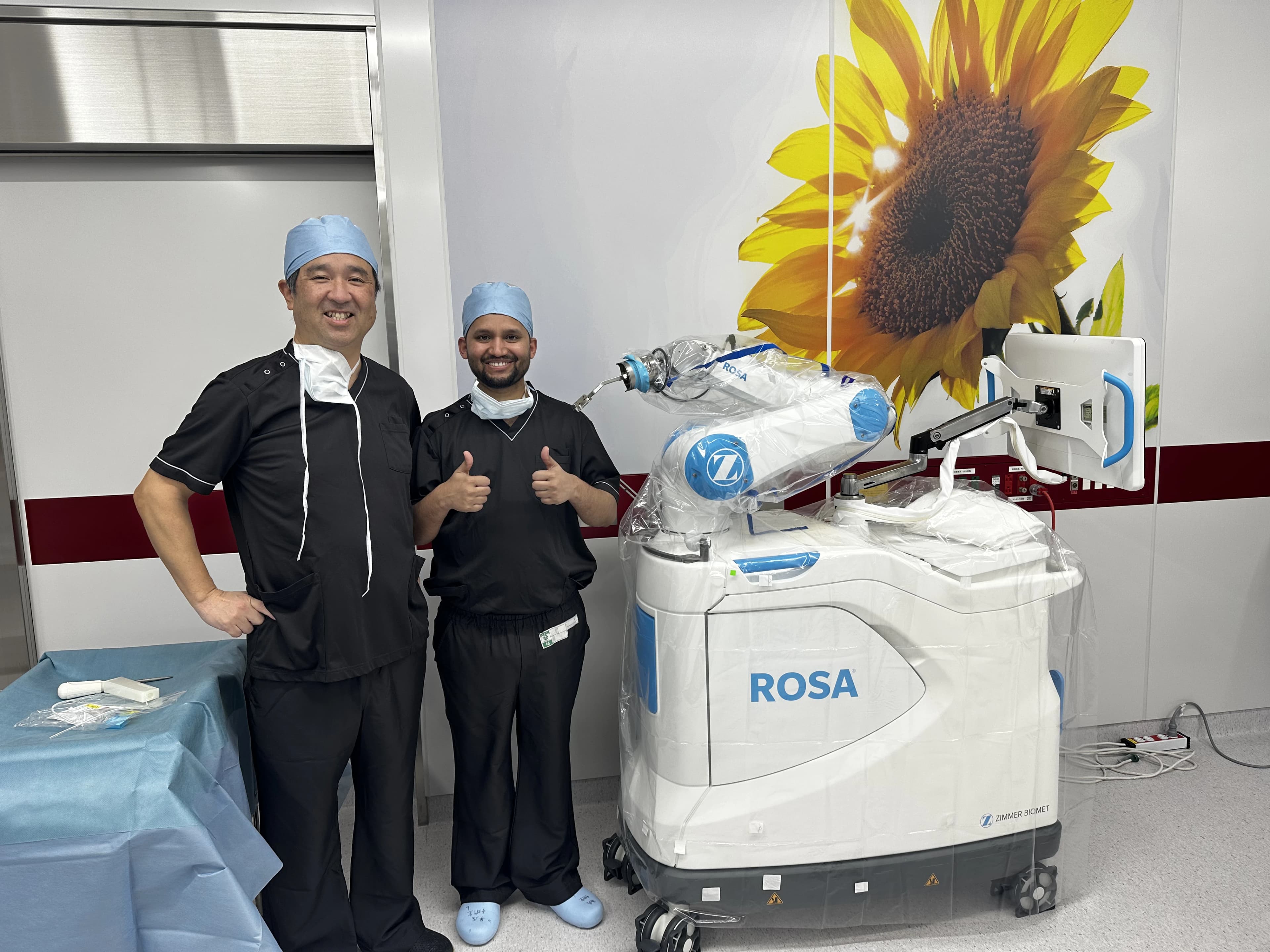
Intraoperative kinematics & 3D navigation alongside Japanese surgical faculty.
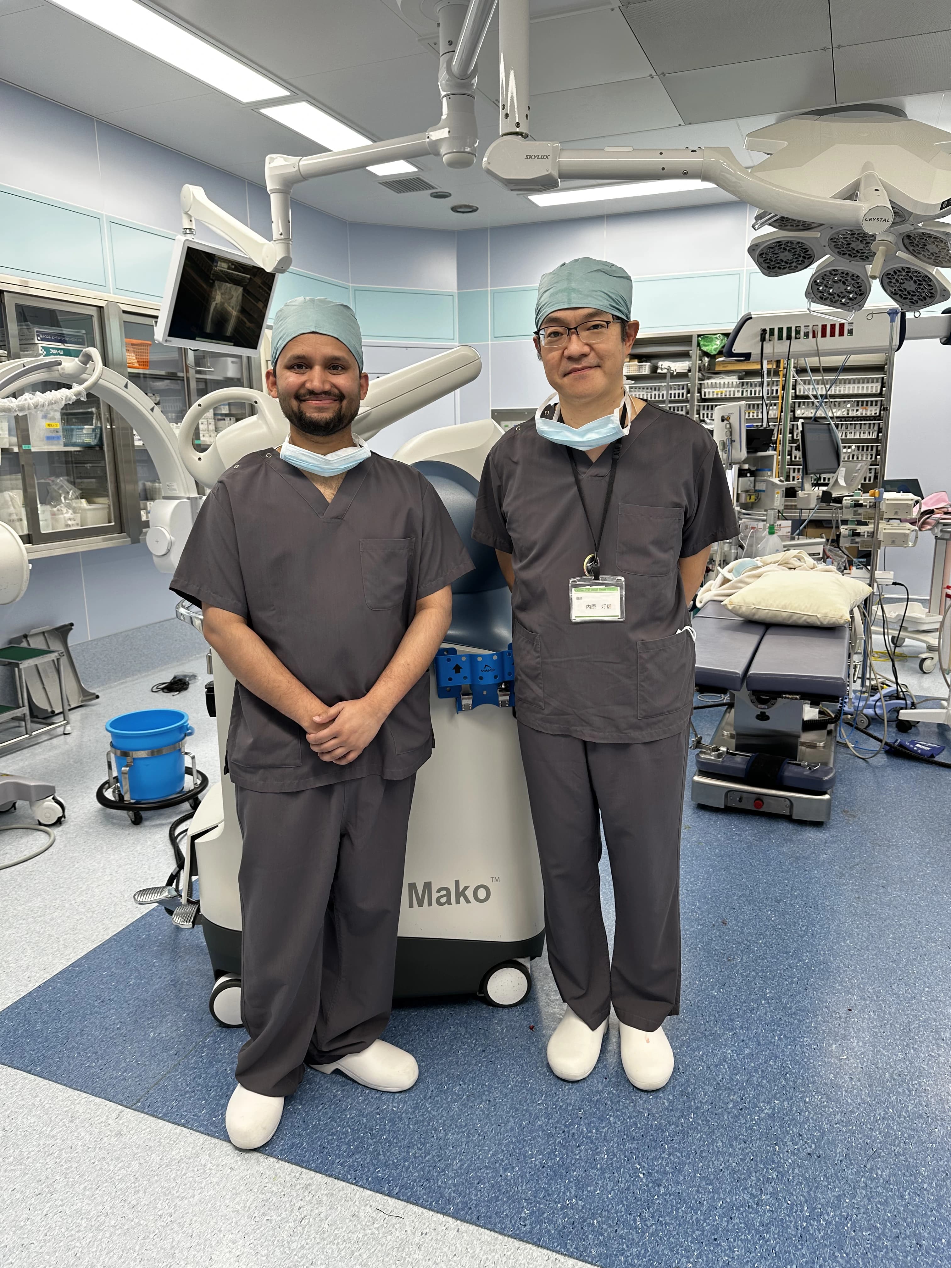
International fellowship-level precision applied to patient care in Patna.
Frequently Asked Questions on Foot & Ankle Fractures
The ankle joint is an extremely precise mortise. Even a 1-mm shift in bone position causes abnormal pressure points that wear away joint cartilage rapidly. Accurate surgical alignment preserves your natural joint for life.