
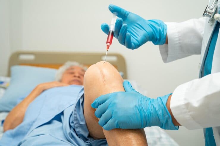

Understanding Cartilage Restoration Procedures & Biological Resurfacing
Focal chondral defects—craters in the smooth articular cartilage of the knee, ankle, or hip—typically result from sports injuries, impacts, or osteochondritis dissecans (OCD). Because articular cartilage does not heal spontaneously, these defects create localized stress concentrations that rapidly degenerate into bone-on-bone arthritis if neglected. Cartilage restoration techniques implant cellular scaffolds or living osteochondral cylinders to resurface the defect.
The Biological Joint-Sparing Philosophy
By correcting mechanical weight-bearing malalignment through precision osteotomy and cartilage preservation, the load is shifted off damaged cartilage onto healthy bone—preserving your natural joint for years to come.
When Should You Consider Consultation for Cartilage Restoration?
Specialist intervention is considered when joint pain or functional limitation significantly disrupts your daily life and conservative measures have been exhausted. Check if you experience any of the following:
Localized joint pain and persistent swelling after athletic pivoting.
Mechanical catching, clicking, or sudden locking sensation during knee flexion.
MRI scan revealing a discrete focal cartilage lesion (Grade III / IV defect).
Young or middle-aged active individual seeking biological alternatives to joint replacement.
Activity-related swelling limiting sports or exercise routines.

Dr. Vijay Kumar
Verified SpecialistSenior Consultant Orthopaedic, Robotic Joint Replacement & Complex Articular Trauma Surgeon
“My surgical philosophy centers on biological preservation, anatomical restoration, and rapid functional recovery. By tailoring the intervention to each patient's unique anatomy and lifestyle, we achieve lasting pain relief and active mobility.”
The 4-Stage Precision Clinical Workflow
Full-Leg Weight-Bearing Alignment Analysis
4K arthroscopic sizing of the defect footprint and vertical shoulder preparation.
Precision Biplanar Osteotomy Planning
Preparation of the subchondral bone bed with micro-drilling.
Rigid Locking Plate Stabilization
Implantation of cellular matrix scaffolds or press-fit osteochondral autografts.
Protected Unloading & Gradual Rehabilitation
Application of biological fibrin sealant.
Stage 05: Joint Preservation Stage
Early protected continuous passive motion protocol.
Treatment Modalities Offered
Dr. Vijay Kumar practices conservative joint-sparing protocols first, proceeding to advanced surgical intervention only when indicated:
Non-Surgical & Joint Sparing
- Unloader knee bracing to eliminate contact pressure
- Platelet-Rich Plasma (PRP) intra-articular injections
- Low-impact cycling and aquatic therapy
- Neuromuscular kinetic chain conditioning
Specialised Surgical Procedures
- Autologous Matrix-Induced Chondrogenesis (AMIC)
- Osteochondral Autograft Transfer (OATS / Mosaicplasty)
- Subchondral Bone Marrow Stimulation with Collagen Membrane
- Removal of Loose Bodies with Chondral Bed Debridement
Recovery Timeline & Mobility Milestones
Weeks 1–4: CPM motion, touch-down weight bearing.
Weeks 6–8: Transition to full weight bearing and stationary cycling.
Months 3–5: Progressive muscular conditioning and swimming.
Months 6–9: High-level agility drills and clearance for running.
International Fellowship & Surgical Suite Records
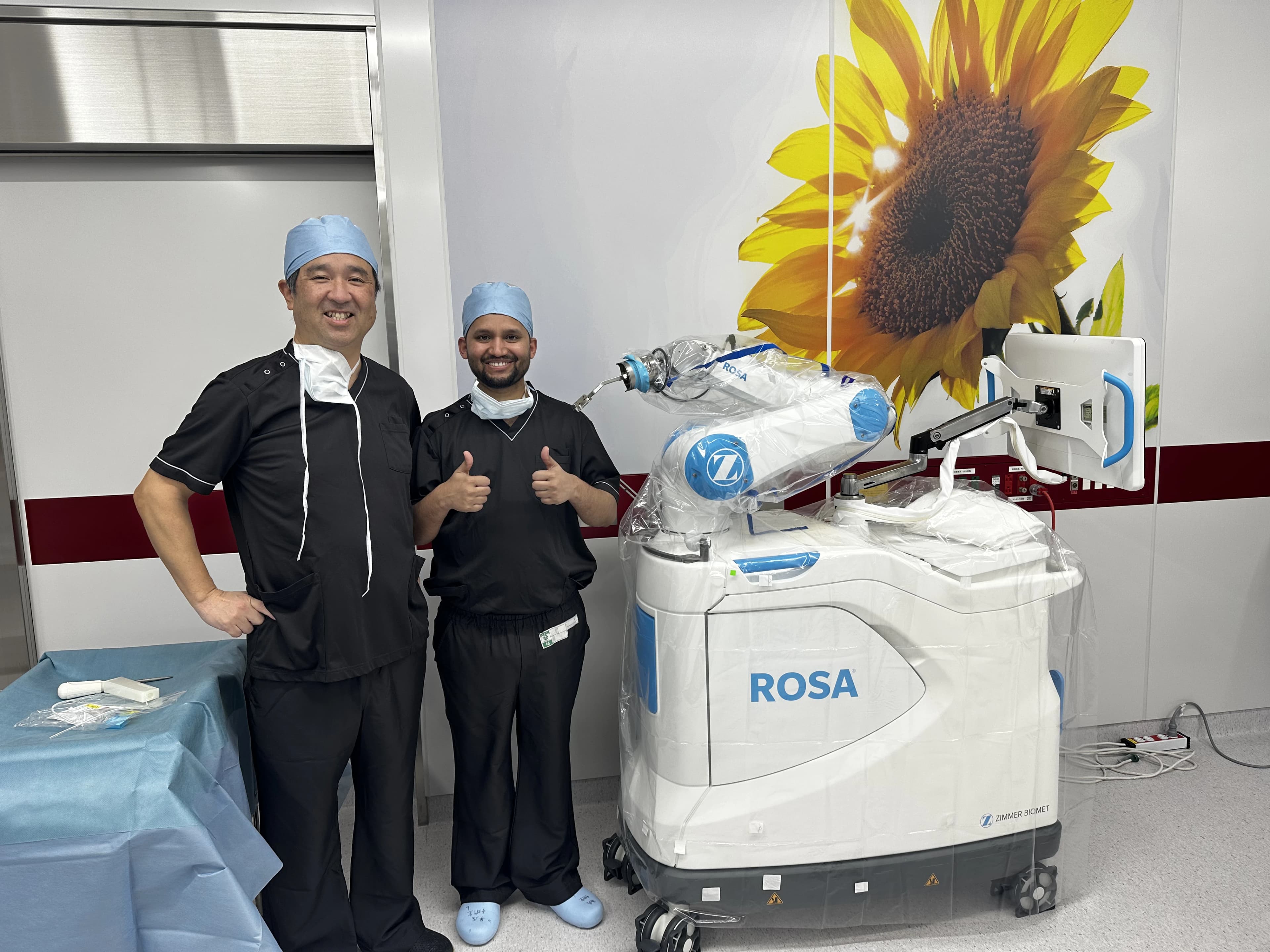
Intraoperative kinematics & 3D navigation alongside Japanese surgical faculty.
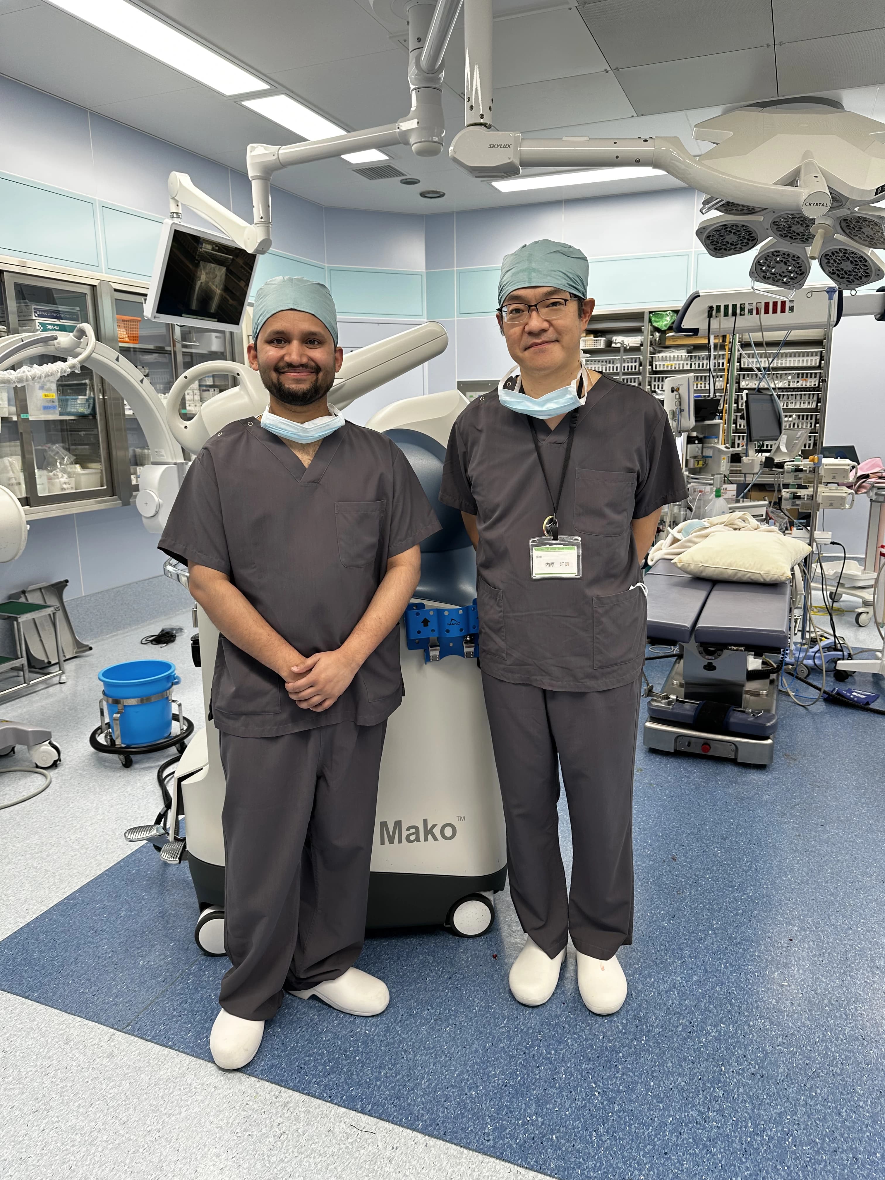
International fellowship-level precision applied to patient care in Patna.
Frequently Asked Questions on Cartilage Restoration
Cartilage restoration is a biological procedure that repairs living tissue using your own cartilage or cellular scaffolds, preserving all your native bones and ligaments.